 1800 605-5127
 +61 (0)8 8352 7711
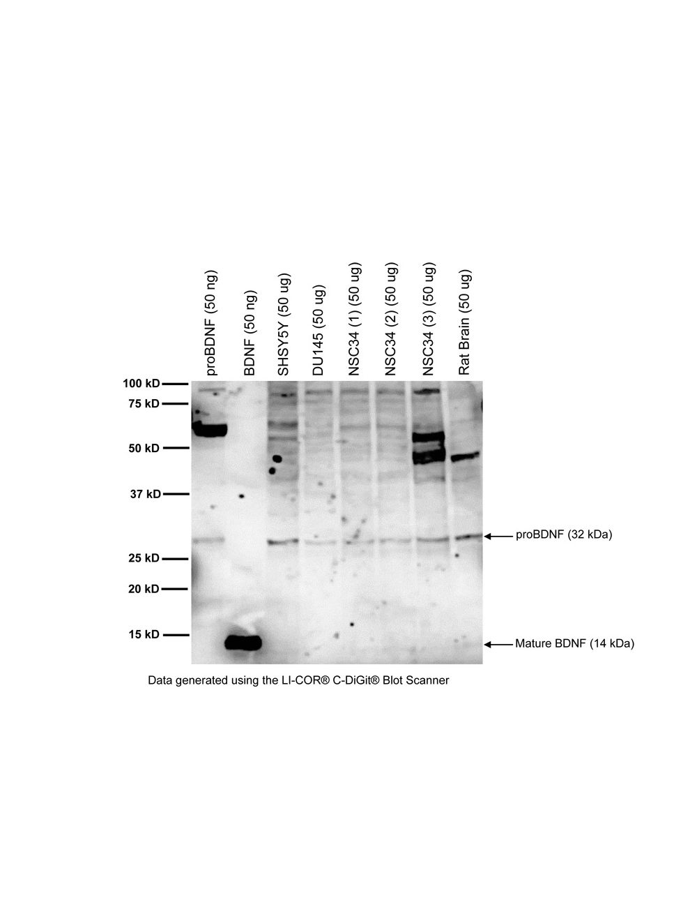

Brain-derived neurotrophic factor (BDNF), Rabbit Polyclonal Antibody
- Product Name Brain-derived neurotrophic factor (BDNF), Rabbit Polyclonal Antibody
-
Product Description
Rabbit anti-Brain-derived neurotrophic factor (BDNF) Polyclonal Antibody (Unconjugated), suitable for WB, IHC-Frozen, ELISA.
- Alternative Names Brain-derived neurotrophic factor; Abrineurin; proBDNF;
- Application(s) ELISA, IHC-Frozen, WB
- Antibody Host Rabbit
- Antibody Type Polyclonal
- Specificity Less than 0.1% cross reactivity with mouse NGF, recombinant human NT3 and NT4/5 has been recorded by dot blot analysis. This antiserum is known to recognise rat, human and human BDNF, and is expected to react with BDNF from other species due to amino acid sequence homology.
- Species Reactivity Human, Mouse, Other Mammals (Predicted), Rat
- Immunogen Description A synthetic peptide (HSDPARRGEL) as a part of human BDNF protein (aa: 129-138) conjugated to KLH has been used as the immunogen. The BDNF protein sequence is highly conserved amongst mammalian species.
- Conjugate Unconjugated
- Purity Description Protein G purified IgG
- Regulatory Status For research use only.
Product Info
-
Product Description
Rabbit anti-Brain-derived neurotrophic factor (BDNF) Polyclonal Antibody (Unconjugated), suitable for WB, IHC-Frozen, ELISA.
- Application(s) ELISA, IHC-Frozen, WB
-
Application Details
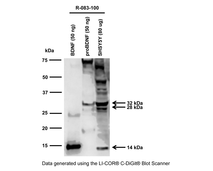
Western Blotting: A concentration of 1-10 µg/mL is recommended for this application. In Western Blotting, this antibody detects multiple BDNF isoforms (14 kDa mature BDNF, 18 kDa isoform, 28 kDa BDNF dimer/truncated BDNF, 32 kDa proBDNF monomer) depending on sample application (human serum, cell lysate, tissue homogenate).
IHC: Antibody works well in immunohistochemistry with the proper fixation, pretreatments and dilution. Formal fixed, paraffin embedded tissue is not recommend. Recommended fixation is Zamboni fixative or light 4% PFA fixation on fixed, frozen tissue. Recommended dilution is 1-10 µg/mL for immunohistochemistry at 4 degrees centigrade for 2-48 hours. ELISA: 1-10 µg/mL capture/detection.
Biosensis recommends optimal dilutions/concentrations should be determined by the end user. - Target Brain-derived neurotrophic factor (BDNF)
- Specificity Less than 0.1% cross reactivity with mouse NGF, recombinant human NT3 and NT4/5 has been recorded by dot blot analysis. This antiserum is known to recognise rat, human and human BDNF, and is expected to react with BDNF from other species due to amino acid sequence homology.
- Target Host Species Human
- Species Reactivity Human, Mouse, Other Mammals (Predicted), Rat
- Antibody Host Rabbit
- Antibody Type Polyclonal
- Antibody Isotype IgG
- Conjugate Unconjugated
- Immunogen Description A synthetic peptide (HSDPARRGEL) as a part of human BDNF protein (aa: 129-138) conjugated to KLH has been used as the immunogen. The BDNF protein sequence is highly conserved amongst mammalian species.
- Purity Description Protein G purified IgG
- Format Lyophilized from PBS, pH 7.4, without preservatives.
- Reconstitution Instructions Spin vial briefly before opening. Reconstitute in 500 µL sterile-filtered ultrapure water, pH 7.2-7.6. Centrifuge to remove any insoluble material.
- Storage Instructions After reconstitution keep aliquots at -20°C for a higher stability, and at 2-8°C with an appropriate antibacterial agent. Glycerol (1:1) may be added for an additional stability. Avoid repetitive freeze/thaw cycles.
- Batch Number Please see item label.
- Expiration Date 12 months after date of receipt (unopened vial).
- Alternative Names Brain-derived neurotrophic factor; Abrineurin; proBDNF;
- Uniprot Number P23560
- Uniprot Number/Name P23560 (BDNF_HUMAN)
-
Scientific Background
BDNF belongs to the neurotrophin family and regulates the survival and differentiation of neurons during development. The alterations in BDNF expression induced by various kinds of brain insult including stress, ischemia, seizure activity and hypoglycemia, may contribute to some pathologies such as depression, epilepsy, Alzheimer, and Parkinson disease. Microglia release BDNF that may contribute to neuroinflammation and neuropathic pain.
FUNCTION: Promotes the survival of neuronal populations that are all located either in the central nervous system or directly connected to it. Major regulator of synaptic transmission and plasticity at adult synapses in many regions of the CNS. The versatility of BDNF is emphasized by its contribution to a range of adaptive neuronal responses including long-term potentiation (LTP), long-term depression (LTD), certain forms of short-term synaptic plasticity, as well as homeostatic regulation of intrinsic neuronal excitability. SUBUNIT: Monomers and homodimers. Binds to NTRK2/TRKB. SUBCELLULAR LOCATION: Secreted protein. POst translation modification: Converted into mature BDNF by plasmin (PLG). SIMILARITY: Belongs to the NGF-beta family. - Shipping Temperature 25°C (ambient)
- UNSPSC CODE 41116161
- Regulatory Status For research use only.
Specifications


- Specific References Feron F et al (2008) Neurotrophin expression in the adult olfactory epithelium. Brain Res. 1196:13-21 Application: IHC; Species: Rat
-
General References
A Acheson et al (1995) Nat. 74: 450-3
Q Yan et al (1996) Neurosci. 74: 945-53
XF Zhou, et al (2004) Crit Rev Neurobiol 16, 43-9
I Tapia-Arancibia et al (2002) Brain Res Brain Res Rev 40, 240-9
Barde Y. A. et al (1989) EMBO J. 1: 549
Conner J et al. (1997) J. Neurosci. 17: 2295
JA Coull et al (2013) J Neuroinflammation. Jan 30;10:16.