1800 605-5127
 +61 (0)8 8352 7711
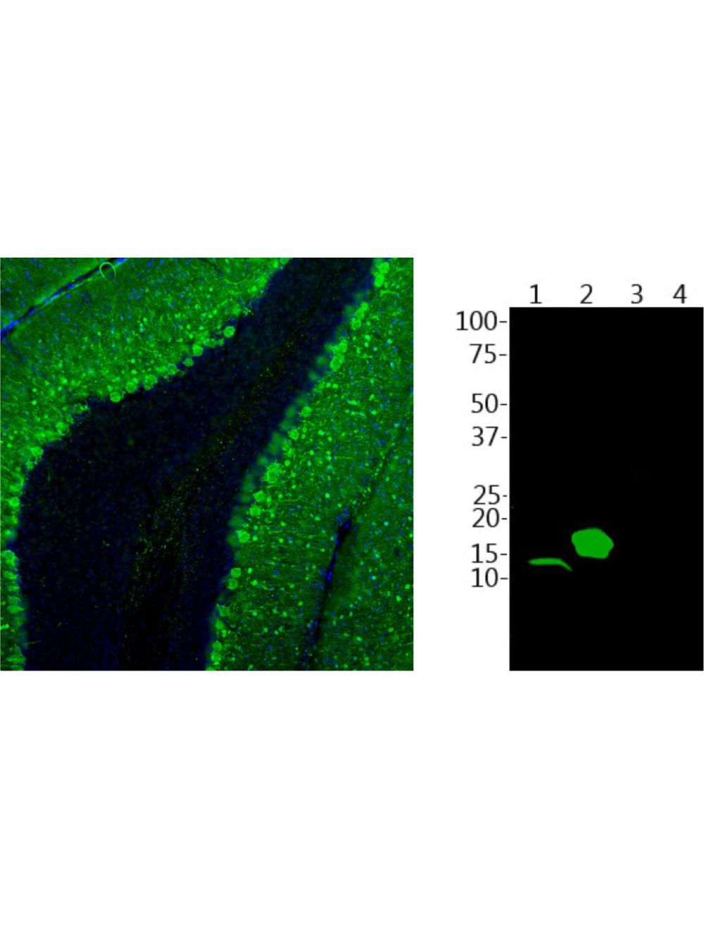

Parvalbumin, Chicken Polyclonal Antibody
- Product Name Parvalbumin, Chicken Polyclonal Antibody
- Product Description Chicken anti-Parvalbumin Polyclonal Antibody (Unconjugated), suitable for WB, IHC-Frozen.
- Alternative Names PVALB; Parvalbumin;
- Application(s) IHC-Frozen, WB
- Antibody Host Chicken
- Antibody Type Polyclonal
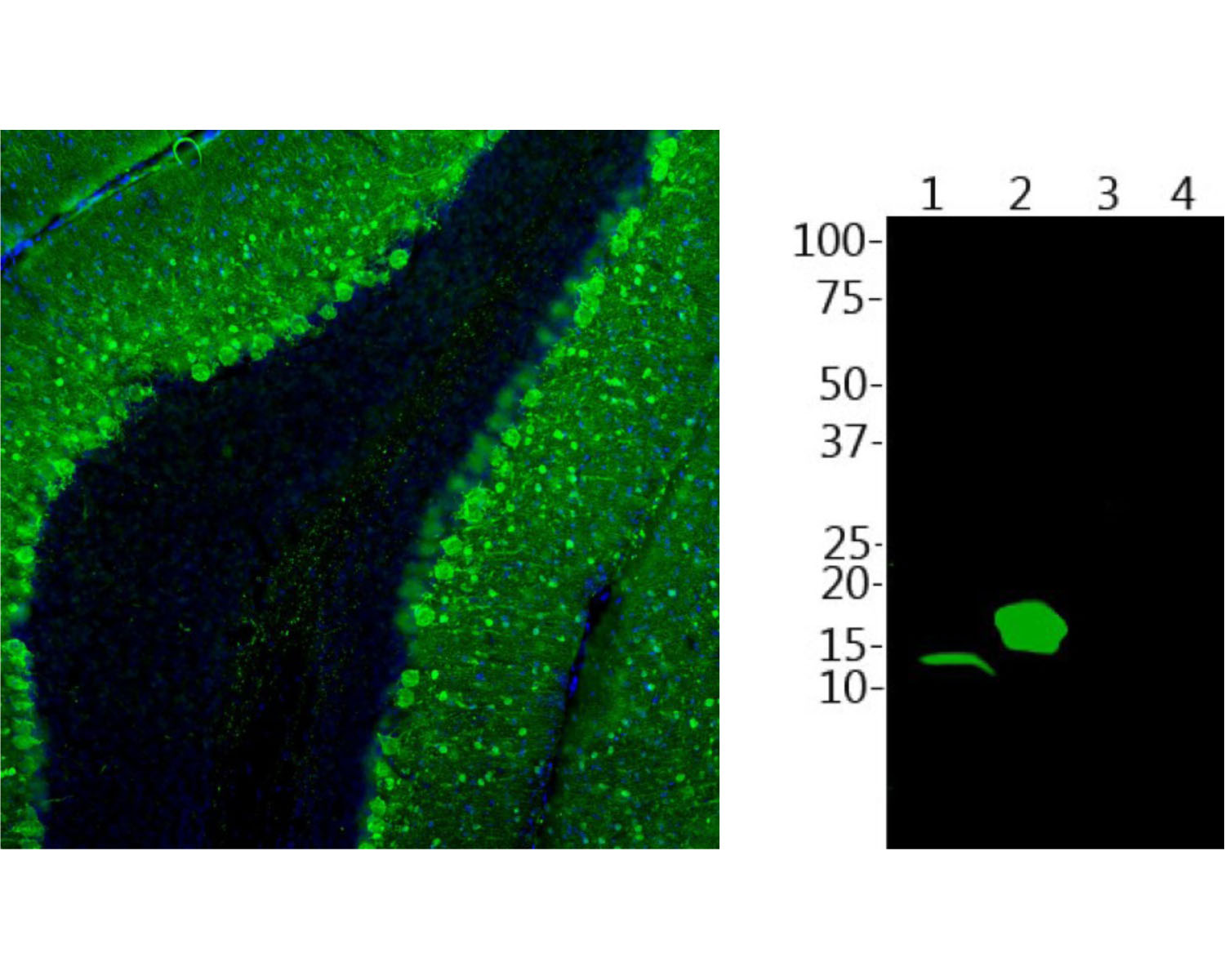
- Specificity Human, reacts with Human, Rat, Mouse. Antibody is specific for parvalbumin and does not recognize closely related proteins calretinin and calbindin as determined by Western Blotting.
- Species Reactivity Human, Mouse, Rat
- Immunogen Description Full-length recombinant human protein
- Conjugate Unconjugated
- Purity Description IgY fraction
- Regulatory Status For research use only.
Product Info
- Product Description Chicken anti-Parvalbumin Polyclonal Antibody (Unconjugated), suitable for WB, IHC-Frozen.
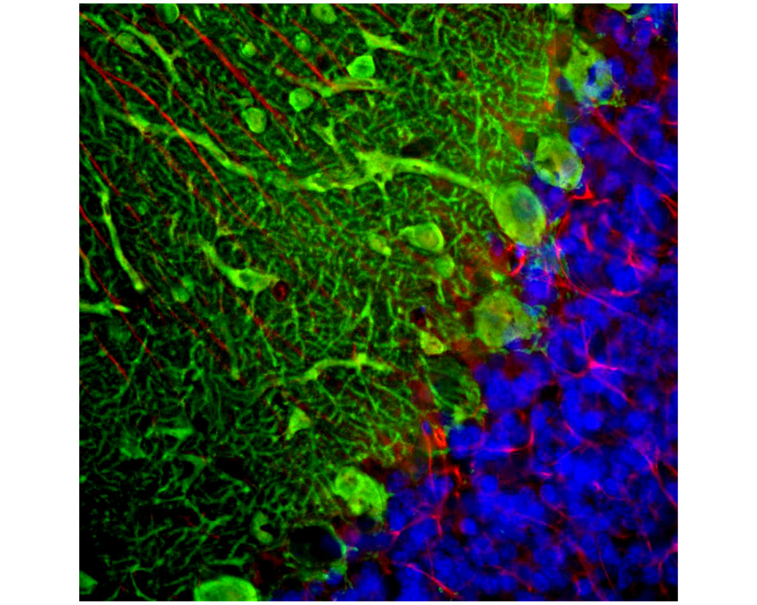
- Application(s) IHC-Frozen, WB
- Application Details Western blotting (1:1,000-1:5,000) and Immunohistochemistry (1:1,000-1:5,000). Note that this antibody does not recognize parvalbumin in rat or mouse brain homogenates on western blots. Biosensis recommends optimal dilutions/concentrations should be determined by the end user.
- Target Parvalbumin
- Specificity Human, reacts with Human, Rat, Mouse. Antibody is specific for parvalbumin and does not recognize closely related proteins calretinin and calbindin as determined by Western Blotting.
- Target Host Species Human
- Species Reactivity Human, Mouse, Rat
- Antibody Host Chicken
- Antibody Type Polyclonal
- Antibody Isotype IgY
- Conjugate Unconjugated
- Immunogen Description Full-length recombinant human protein
- Purity Description IgY fraction
- Format Lyophilized IgY preparation, with sodium azide.
- Reconstitution Instructions Spin vial briefly before opening. Reconstitute with 50 µL sterile-filtered, ultrapure water. Centrifuge to remove any insoluble material.
- Storage Instructions Store lyophilized antibody at 2-8°C. After reconstitution divide into aliquots and store at -20°C for long-term storage. Store at 2-8°C short-term (up to 4 weeks) with an appropriate antibacterial agent. Avoid repetitive freeze/thaw cycles.
- Batch Number Please see item label.
- Expiration Date 12 months after date of receipt (unopened vial).
- Alternative Names PVALB; Parvalbumin;
- Uniprot Number P20472
- Uniprot Number/Name P20472 (PRVA_HUMAN)
- Scientific Background In muscle, parvalbumin is thought to be involved in relaxation after contraction. It binds two calcium ions. Ref: uniprot.org
- Shipping Temperature 25°C (ambient)
- UNSPSC CODE 41116161
- Regulatory Status For research use only.