 1800 605-5127
 +61 (0)8 8352 7711
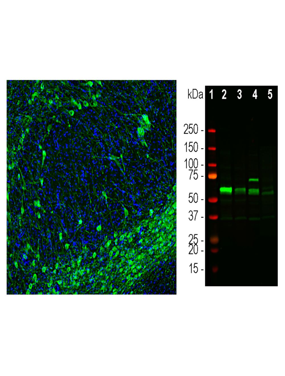

Tyrosine Hydroxylase (TH), Chicken Polyclonal Antibody
As low as
US$317.00
Only %1 left
Catalog Number
C-2126
- Product Name Tyrosine Hydroxylase (TH), Chicken Polyclonal Antibody
- Product Description Chicken anti-Tyrosine Hydroxylase (TH), Polyclonal Antibody (Unconjugated), suitable for WB, IHC-Frozen, ICC, FC
- Alternative Names TH; T-H; tyrosine 3-monooxygenase; monooxygenase; oxidoreductase
- Application(s) FC, ICC, IHC-Frozen, WB
- Antibody Host Chicken
- Antibody Type Polyclonal
- Specificity Human. Species cross-reactivity includes rat and mouse.
- Species Reactivity Human, Mouse, Rat
- Immunogen Description The antibody has been made against the full length recombinant human TH (524 amino acid sequence) expressed in and purified from E. Coli
- Conjugate Unconjugated
- Purity Description Affinity-purified
- Regulatory Status For research use only.
Product Info
- Product Description Chicken anti-Tyrosine Hydroxylase (TH), Polyclonal Antibody (Unconjugated), suitable for WB, IHC-Frozen, ICC, FC
-
Related Products
Tyrosine Hydroxylase (TH), Rabbit Polyclonal Antibody
- Application(s) FC, ICC, IHC-Frozen, WB
- Application Details Western Blotting (WB), Immunocytochemistry (ICC), Immunohistochemistry (IHC). A dilution of 1:5,000 - 1:10,000 is recommended for WB. A dilution of 1:100 - 1:500 is recommended for IC and IH. Flow Cytometry at 20 µg/mL. Biosensis recommends optimal dilutions/concentrations should be determined by the end user.
- Target Tyrosine Hydroxylase (TH); Tyrosine 3-monooxyegnase
- Specificity Human. Species cross-reactivity includes rat and mouse.
- Target Host Species Human
- Species Reactivity Human, Mouse, Rat
- Antibody Host Chicken
- Antibody Type Polyclonal
- Antibody Isotype IgY
- Conjugate Unconjugated
- Immunogen Description The antibody has been made against the full length recombinant human TH (524 amino acid sequence) expressed in and purified from E. Coli
- Purity Description Affinity-purified
- Format Lyophilized from PBS buffer pH 7.2-7.6 with 0.1% trehalose, and sodium azide
- Reconstitution Instructions Spin vial briefly before opening. Reconstitute with 100 µL sterile-filtered, ultrapure water to achieve a 1 mg/mL concentration. Centrifuge to remove any insoluble material.
- Storage Instructions After reconstitution of lyophilized antibody, aliquot and store at -20°C for a higher stability. Avoid freeze-thaw cycles. Store at 4°C for up to one month for short term storage and frequent use.
- Batch Number Please see item label.
- Expiration Date 12 months after date of receipt (unopened vial).
- Alternative Names TH; T-H; tyrosine 3-monooxygenase; monooxygenase; oxidoreductase
- Uniprot Number P07101
- Uniprot Number/Name P07101 (TY3H_HUMAN)
- Scientific Background Catalyzes the conversion of L-tyrosine to L-dihydroxyphenylalanine (L-Dopa), the rate-limiting step in the biosynthesis of cathecolamines, dopamine, noradrenaline, and adrenaline. Uses tetrahydrobiopterin and molecular oxygen to convert tyrosine to L-Dopa. In addition to tyrosine, is able to catalyze the hydroxylation of phenylalanine and tryptophan with lower specificity (By similarity). Positively regulates the regression of retinal hyaloid vessels during postnatal development (By similarity). (Ref: uniprot.org)
- Shipping Temperature 25°C (ambient)
- UNSPSC CODE 41116161
- Regulatory Status For research use only.
