 1800 605-5127
 +61 (0)8 8352 7711
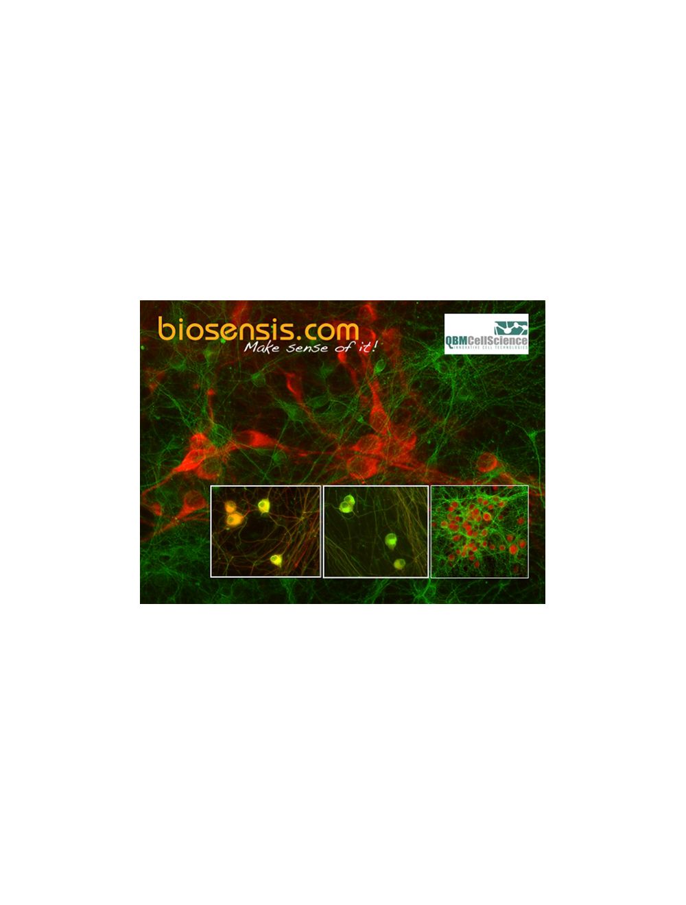

Peripherin, Chicken Polyclonal Antibody
- Product Name Peripherin, Chicken Polyclonal Antibody
- Product Description Chicken anti-Peripherin Polyclonal Antibody (Unconjugated), suitable for WB, IHC-Frozen, IHC-Paraffin-embedded, ICC.
- Alternative Names Peripherin; Prph; Prph1;
- Application(s) ICC, IHC-Frozen, IHC-Paraffin-embedded, WB
- Antibody Host Chicken
- Antibody Type Polyclonal
- Specificity The specificity of this antibody has been confirmed by WB. This antibody detects ~57 kDa Peripherin protein. A suitable control tissue is rat spinal cord or peripheral nerve homogenate. Hu, Rat, Ms, Fel, and other mammals
- Species Reactivity Cat, Human, Mouse, Other Mammals, Rat
- Immunogen Description Recombinant full length rat Peripherin protein expressed in and purified from E.coli
- Conjugate Unconjugated
- Purity Description IgY
- Regulatory Status For research use only.
Product Info
- Product Description Chicken anti-Peripherin Polyclonal Antibody (Unconjugated), suitable for WB, IHC-Frozen, IHC-Paraffin-embedded, ICC.
- Application(s) ICC, IHC-Frozen, IHC-Paraffin-embedded, WB
- Application Details Western Blotting (WB) and Immunocytochemistry (ICC). A dilution of 1:5,000 - 1:10,000 is recommended for WB. A dilution of 1:500-1,000 is recommended for IC. Biosensis recommends optimal dilutions/concentrations should be determined by the end user.
- Target Peripherin
- Specificity The specificity of this antibody has been confirmed by WB. This antibody detects ~57 kDa Peripherin protein. A suitable control tissue is rat spinal cord or peripheral nerve homogenate. Hu, Rat, Ms, Fel, and other mammals
- Target Host Species Rat
- Species Reactivity Cat, Human, Mouse, Other Mammals, Rat
- Antibody Host Chicken
- Antibody Type Polyclonal
- Antibody Isotype IgY
- Conjugate Unconjugated
- Immunogen Description Recombinant full length rat Peripherin protein expressed in and purified from E.coli
- Purity Description IgY
- Format Lyophilized IgY preparation, with sodium azide.
- Reconstitution Instructions Spin vial briefly before opening. Reconstitute with 50 µL sterile-filtered, ultrapure water. Centrifuge to remove any insoluble material.
- Storage Instructions After reconstitution of lyophilized antibody, aliquot and store at -20°C for a higher stability. Avoid freeze-thaw cycles.
- Batch Number Please see item label.
- Expiration Date 12 months after date of receipt (unopened vial).
- Alternative Names Peripherin; Prph; Prph1;
- Uniprot Number P21807
- Uniprot Number/Name P21807 (PERI_RAT)
- Scientific Background Peripherin is a class-III neuronal intermediate filament protein found in certain classes of neuron, most of which are located in the peripheral nervous system.
- Shipping Temperature 25°C (ambient)
- UNSPSC CODE 41116161
- Regulatory Status For research use only.
Specifications


-
Specific References
Sekerkova G. et al (2008) Espin actin-cytoskeletal proteins are in rat type I spiral ganglion neurons and include splice-isoforms with a functional nuclear localization signal. J Comp Neurol. 2008 Aug 20;509(6):661-76.